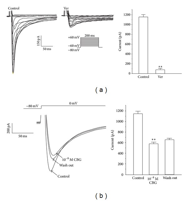
Figure 2.

Ver (0.1 mM) completely blocks I Ca-L in rat ventricular myocytes. (a) Representative I Ca-L recordings according to the steady-state activation protocol before and after application of Ver. Summary data of I Ca-L before and after application of Ver. (b) I Ca-Lwas recorded under control conditions, during exposure to 10−8 M CBG and during wash out. Data are means ± S.E.M. (n = 6 cells). **P < 0.01, versus control.
