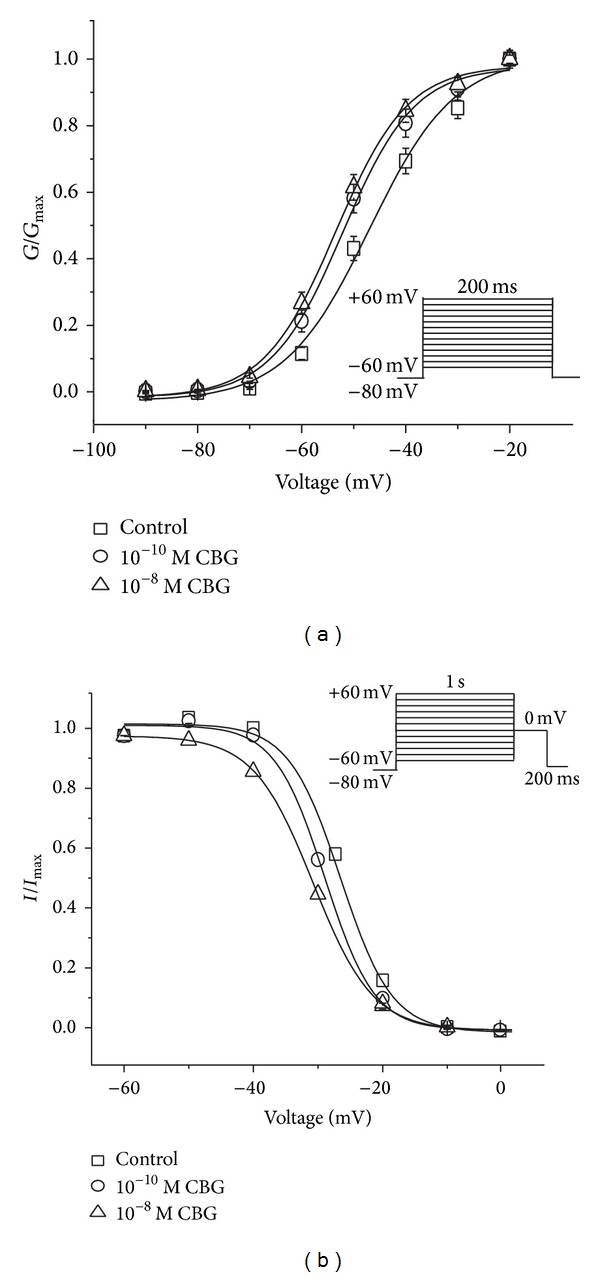
Figure 6.

Effects of CBG on voltage-gated LTCCs in isolated rat left ventricular myocytes. (a) The steady-state activation of LTCCs shifted left by application of 10−10 and 10−8 M CBG. Tail currents were elicited by depolarization to −60 mV after 200 ms test pulses from −60 to 0 mV in increments of 10 mV. (b) CBG shifted the steady-state inactivation curve of LTCCs to the hyperpolarizing direction. The voltage protocol included double pulses consisting of a 200 ms test pulse to 0 mV following a 1 s conditioning pulse varying from −60 to 60 mV with 10 mV increments. Data are presented as means ± S.E.M. (n = 8 cells).
