Figure 1.

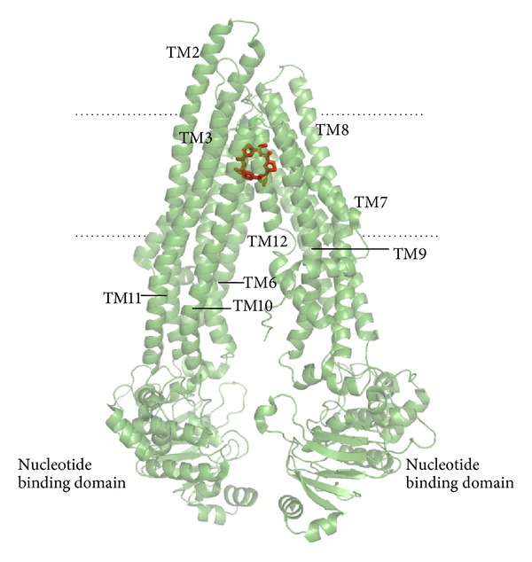
Crystal structure of the complex of mouse P-gp colored in green with its inhibitor in red (pdb code 3G60). Some visible transmembrane domains are labeled (TM). Dashed horizontal lines approximate the region of the lipid bilayer.

Crystal structure of the complex of mouse P-gp colored in green with its inhibitor in red (pdb code 3G60). Some visible transmembrane domains are labeled (TM). Dashed horizontal lines approximate the region of the lipid bilayer.