
Figure 3.
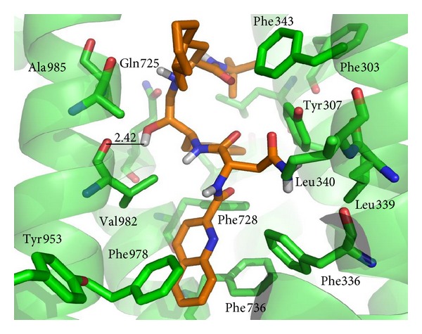
Binding geometry of compound Saquinavir into the P-gp binding pocket predicted by the Autodock docking algorithms (simulation 5). Residues of the binding pocket are highlighted in green. Only the N-bound and O-bound H atoms of the ligand are shown. Carbon atoms of Saquinavir are highlighted in orange. Values of the relevant distances are given in Å.