
Figure 2.
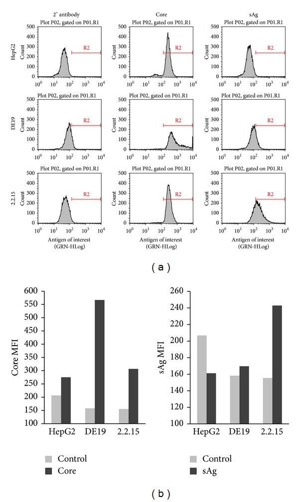
HBV protein expression in infected cell lines. (a) HepG2, DE19 (lacks sAg), and 2.2.15 (complete HBV genome) were harvested, fixed and permeabilized, and stained with HBV specific antibodies directed against core or sAg. (b) Median fluorescent intensity (MFI) derived from the flow plots shown in (a).