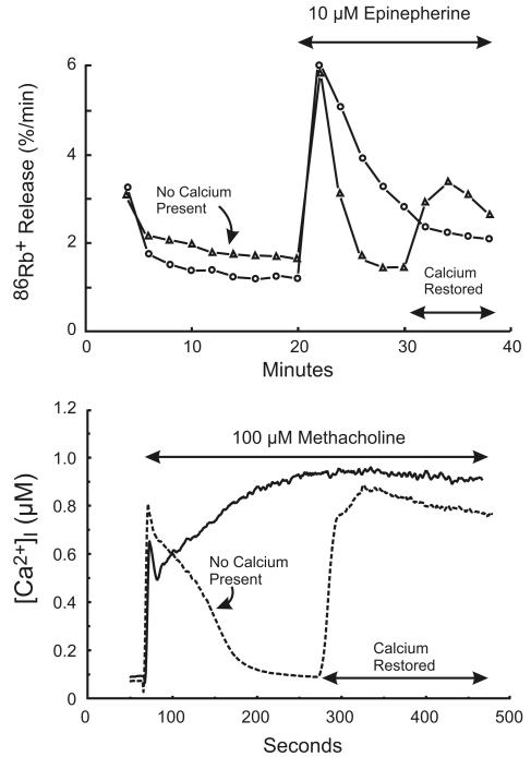
Figure 1. Two phases of Ca2+ signaling in in vitro lacrimal gland preparations.
Top: Changes in intracellular Ca2+ in slices of rat lacrimal gland are inferred from the efflux rate of 86Rb+. Redrawn from data originally presented in [11]. Bottom: Changes in intracellular Ca2+ in mouse lacrimal acinar cells are measured with the Ca2+ indicator, Fura-2. In both cases, in the absence of extracellular Ca2+, the response is transient, and subsequently restored by addition of Ca2+.

