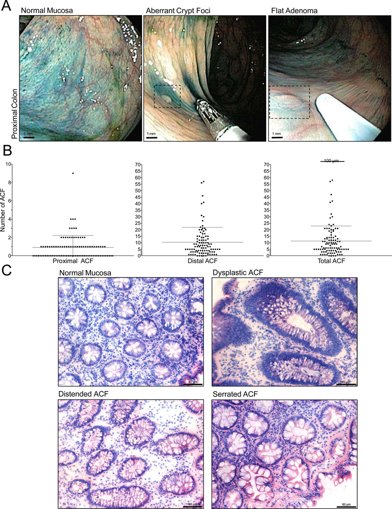
Figure 1. Endoscopic detection of proximal ACF.
A, High-definition magnifying colonoscopy combined with dye-spray reveals diminutive flat lesions of the proximal colorectum, including ACF and flat adenomas. B, Frequency of ACF according to colonic location identified during screening chromoendoscopy. C, Histologic appearance of normal colonic mucosa and ACF procured from the proximal colon. Proximal lesions display the same type of morphologies commonly associated with distal colon ACF, including dysplasia and hyperplasia (distended and serrated) (200× magnification; bar = 60 µm).

