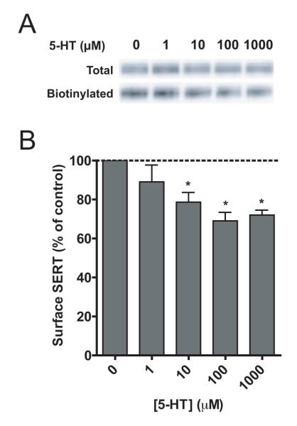
Figure 3.
Biotinylation of surface proteins in HEK293 cells stably expressing c-myc hSERT. Cells are preincubated with or without 5-HT at the indicated concentrations for 30 min at 37 °C. (A) Representative SERT immunoblot of three separate experiments. (B) Quantitive analysis of SERT band densities using Adobe Photoshop. Biotinylated fractions (surface protein) are normalized to total protein and subsequently expressed as percentage of control (preincubation without 5-HT). Data represents the mean (± SEM) of 3 independent experiments. * denotes significant different from control using one-sample t-test (P < 0.05).

