Figure 1.
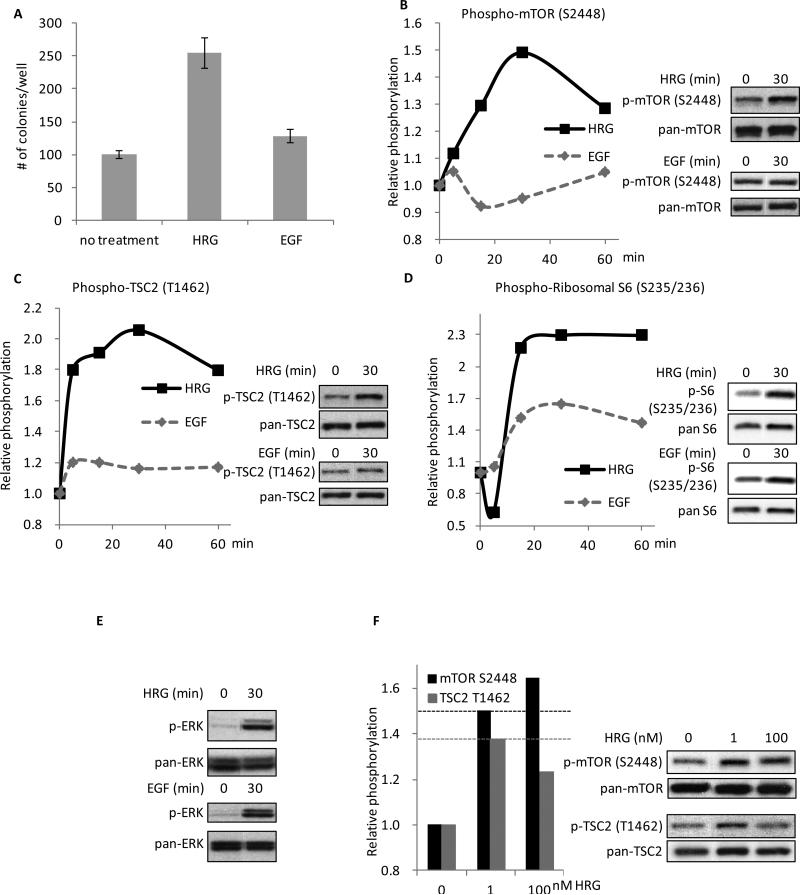
HRG is more effective than EGF at promoting colony formation in SKBR3 cells and in signaling to mTORC1. A, SKBR3 cells were seeded in 0.3% agarose-containing complete medium with the addition of 100 nM HRG or 100 ng/ml EGF. Cells were fed every three days with the growth factor-containing medium and colonies were counted on day 13. The experiment was done in triplicate and the results were averaged and graphed (p-values: Control vs. HRG = 0.0008; Control vs. EGF = 0.0268; HRG vs. EGF = 0.0021). B, SKBR3 cells were serum-starved for 40-48 h followed by 0-60 minutes of treatment with 100 nM HRG or 100 ng/ml EGF. Whole cell lysates were collected and subjected to Western blotting with phospho-mTOR (S2448) and pan-mTOR antibodies. The experiment was performed in triplicate and one representative blot was quantified using ImageJ. Relative intensities of the bands were taken as a ratio of the phospho-protein over total-protein and then plotted against the zero-minute time-point of each individual blot, which was normalized to one. Two time points, 0 and 30 minutes, are shown on the right as an example of the Western blots. C, SKBR3 cells were treated as stated above. Whole cell lysates were collected and subjected to Western blotting with phospho-TSC2 (T1462) and pan-TSC2 antibodies. The blots were quantified as described above. Two time points (0 and 30 minutes) are shown on the right as an example of the Western blots. D, SKBR3 cells were treated as stated above. Whole cell lysates were collected and subjected to Western blotting with phospho-ribosomal S6 (S235/236) and pan-ribosomal S6 antibodies. The blots were quantified as described above. Two time points (0 and 30 minutes) are shown on the right as an example of the Western blots. E, SKBR3 cells were treated as stated above. Whole cell lysates were collected and subjected to Western blotting with phospho-ERK (T202/Y204) and pan-ERK antibodies. Two time points (0 and 30 minutes) are shown as an example of the Western blots. F, SKBR3 cells were serum-starved for 40-48 h followed by 0, 1, or 100 nM HRG stimulation for 30 minutes. Whole cell lysates were collected and subjected to Western blotting with antibodies against phospho-mTOR (S2448), phospho-TSC2 (T1462), pan-mTOR, and pan-TSC2. The blots were quantified as described above.

