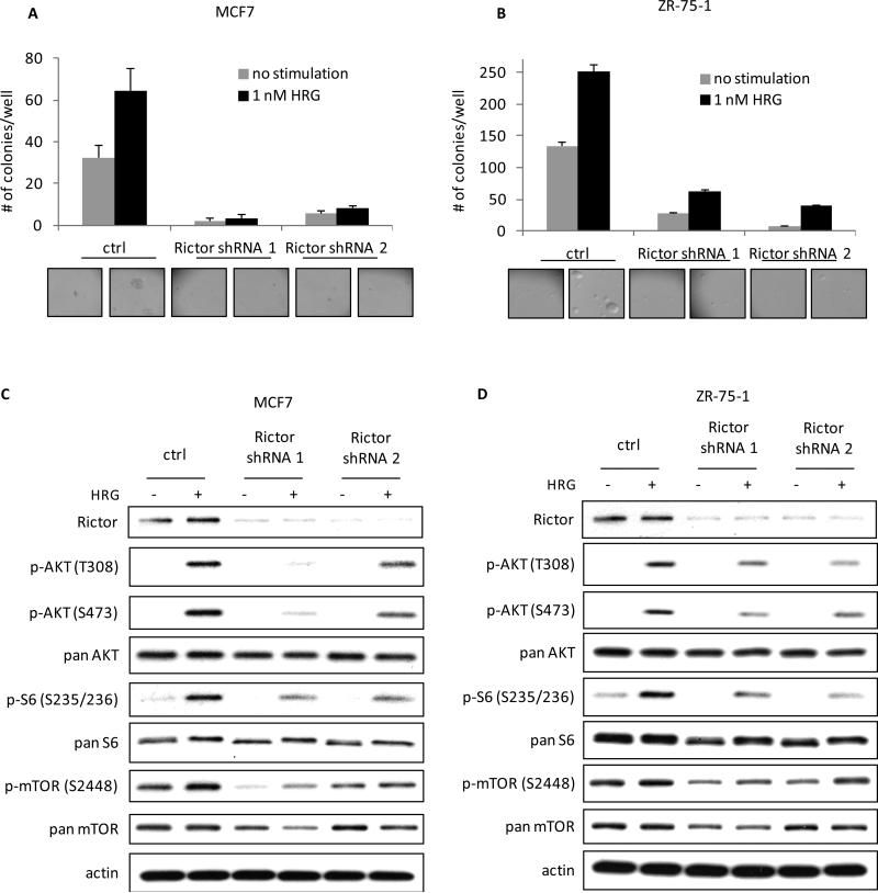
Figure 6.
MCF7 and ZR-75-1 breast cancer cells require signaling to mTORC2 for their ability to form colonies in soft agar and to activate mTORC1 in response to HRG. A, mTORC2 was disrupted in MCF7 cells by infecting the cells with viruses containing control or 2 distinct Rictor shRNAs twice, one day apart, followed by 48 h selection with 2 μg/ml puromycin. Cells were then seeded in 0.3% agarose in complete medium with or without 1 nM HRG. Cells were fed every three days and colonies were counted after 18 days. The experiment was done in triplicate and the results were averaged and graphed. B, mTORC2 was disrupted in ZR-75-1 cells as described in Figure 6A and the cells were then analyzed for their ability to form colonies in soft agar in the presence or absence of 1 nM HRG. C, MCF7 cells were infected and selected as described above. Cells were then serum-starved for 18-24 h, followed by stimulation with 1 nM HRG for 30 minutes. Whole cell lysates were collected and subjected to Western blotting. Blots were probed for Rictor, phospho-AKT (T308), phospho-AKT (S473), phospho-ribosomal S6 (S235/236), phospho-mTOR (S2448), the corresponding antibodies for total protein, and actin. D, ZR-75-1 cells were infected with Rictor shRNAs, selected with 2 μg/ml puromycin and then serum-starved for 18-24 h. After a 30 minute treatment with or without HRG, cells were collected and analyzed by Western blotting as described in Figure 6C.

