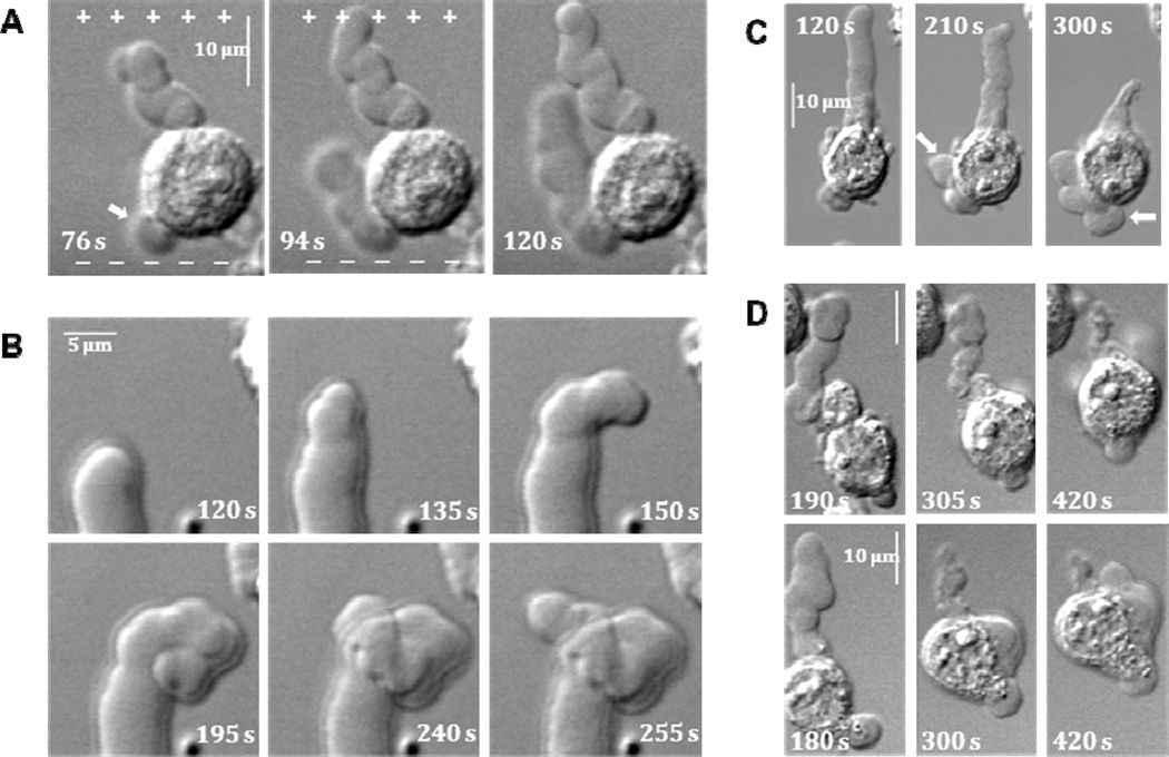
Figure 3.
Distinctive features of PLB formation and lifecycle. For all panels, nsPEF exposure lasts from 0 s to 119 s, and the position of electrodes with respect to cells is the same as in Figs. 1 and 2.
A: nsPEF guides PLB growth towards anode even if PLB starts elsewhere on the cell body. In the cell shown, a bleb that nucleated on the left side of the cell body (arrow, 76 s) later turns and grows towards anode (+).
B : Undirected PLB growth after the end of nsPEF exposure. Sequential images illustrate the loss of directionality of PLB in one of the cells from Fig. 2.
C : The volume of liquid extruded from PLB during its shrinkage following nsPEF exposure is accommodated in secondary blebs (arrows).
D: Examples of cell translocation by PLB contraction. Upper and lower row show two individual cells from different experiments.

