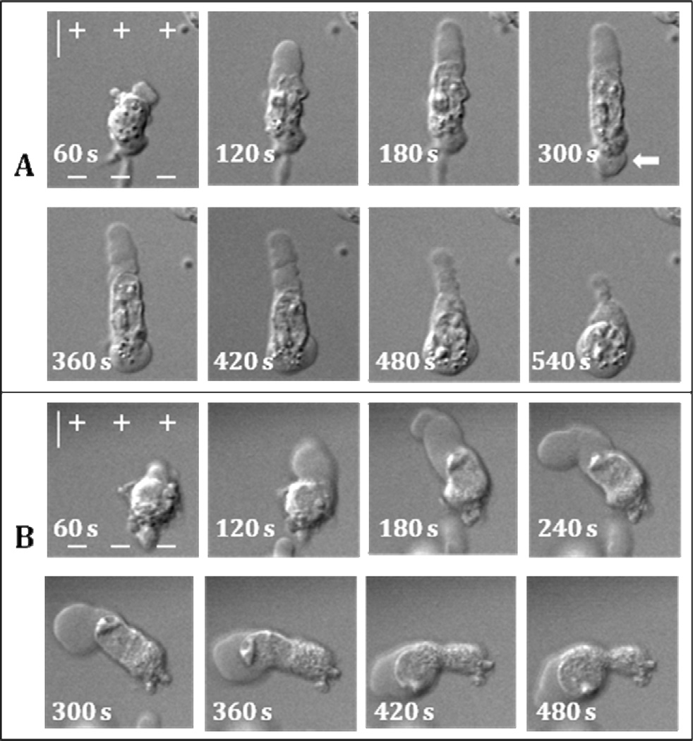
Figure 7.
Destruction of microtubules increases mobility and deformation of nuclei during PLB extension and retraction. NsPEF exposure is from 0 s to 119 s. A and B: two individual cells that show different behavior of cell nucleus after nocodazole treatment. In A, the nucleus gets pulled into PLB but then migrates back and into the secondary bleb on the cathodic cell pole (arrow). In B, nucleus squeezes through PLB neck and enters bleb lumen. The scale is 10 µm. See text and Figs. 1 and 2 for more detail.

