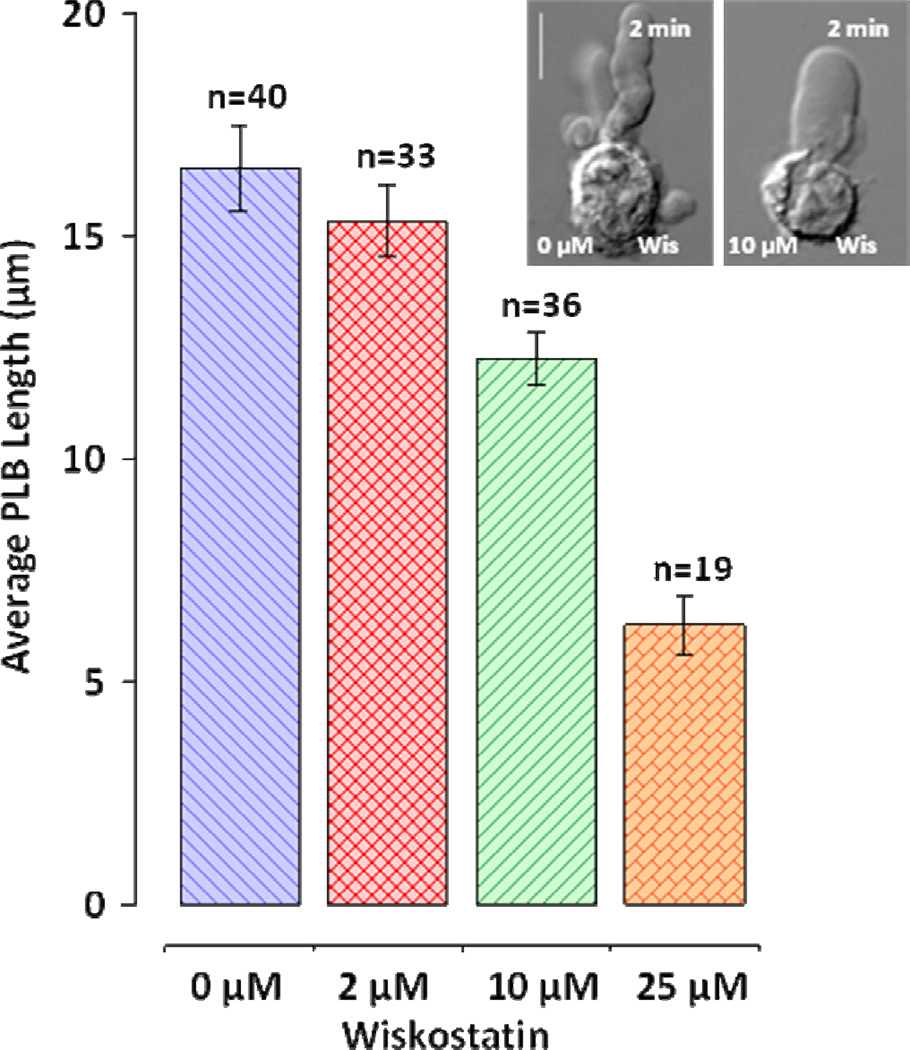
Figure 8.
Inhibition of WASP by wiskostatin inhibits PLB growth. Peak PLB length (mean +/− S.E.) was measured immediately after nsPEF treatment. Cells were placed in the drug-containing buffer immediately prior to nsPEF treatment. Inhibition by 10 µM and 25 µM wiskostatin is significant at p<0.01 (Student’s t-test) as compared to control. The scale bar is 10 µm. The inset shows typical blebs formed in the control buffer and in the presence of 10 µM wiskostatin. Note concurrent thickening and shortening of PLB. See text for more detail.

