
Figure 2.
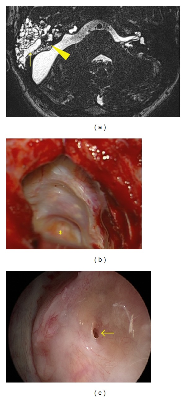
Medial variant of CSF leak. (a) T2W MRI, arrowhead pointing to the fistula; arrow demonstrates CSF filled pneumatic system of temporal bone. (b) Wound revision with identification of fistula in the posterior rim of meatotomy; asterisk shows the closed IAC. (c) Endoscopic view of the fistula.