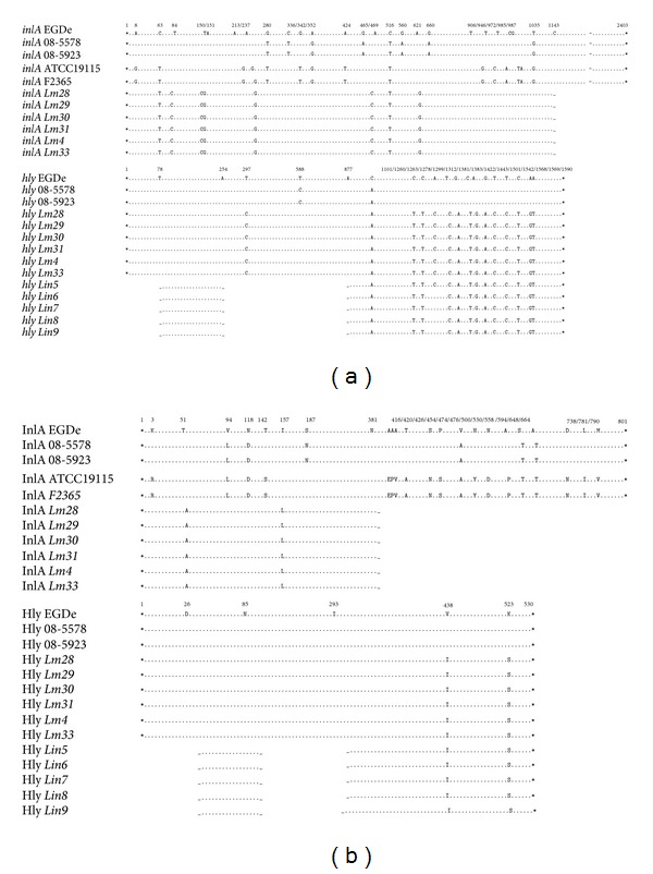
Figure 3.

Nucleotide substitutions detected in inlA and hly (a) and mutations identified in InlA and Hly (b). The Lm28–31, Lm4, Lm33, and Lin5–9 isolates were aligned with L. monocytogenes EGDe and previously described mutant strains. Asterisks indicate the start and stop codons, dots represent identical amino acids, and numbers indicate the positions of the substitutions. Gaps represent the regions that were not amplified.
