
Figure 1.
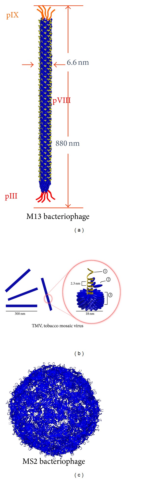
Schematic diagram of various distinct structures of various phages. (a) Long rod structure of M13 bacteriophage with genomic schematic diagrams to show each protein expressed on the M13 phage surfaces. (b) Structure of Tobacco mosaic virus, a rod-like structured plant virus, made of single strand RNA ① and capsid ③ composed of coat ② proteins. (c) Sphere structure of MS2 bacteriophage.