
Figure 1.
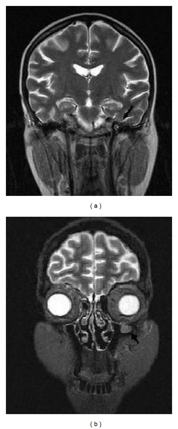
The cranial and orbital sagittal T2-weighted magnetic resonance image revealing a thickening of the third division of trigeminal nerve (a) and a soft tissue development of one centimeter diameter in the roof of left maxillary sinus (b).
Official websites use .gov
A
.gov website belongs to an official
government organization in the United States.
Secure .gov websites use HTTPS
A lock (
) or https:// means you've safely
connected to the .gov website. Share sensitive
information only on official, secure websites.
The cranial and orbital sagittal T2-weighted magnetic resonance image revealing a thickening of the third division of trigeminal nerve (a) and a soft tissue development of one centimeter diameter in the roof of left maxillary sinus (b).