Fig. 3.

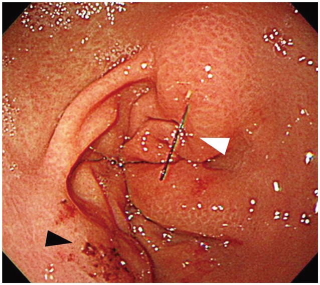
Endoscopic findings. A needle is embedded in the posterior wall of the antrum with edematous mucosal change around the needle (white arrowhead). Mucosal erosion and inflammation are seen on the opposite side of the needle (black arrowhead).

Endoscopic findings. A needle is embedded in the posterior wall of the antrum with edematous mucosal change around the needle (white arrowhead). Mucosal erosion and inflammation are seen on the opposite side of the needle (black arrowhead).