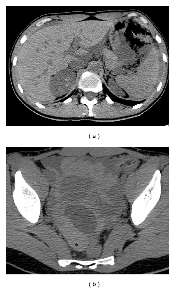
Figure 2.

Unenhanced CT axial images (a, b) show diffuse peritoneal effusion in abdomen with different attenuation values that progressively increase from the upper abdomen (a) to the pelvis (b) where it becomes strongly hyperdense (60–65 HU,*) for the presence of blood component. (b) Low-density cyst is appreciable in the right adnexal area.
