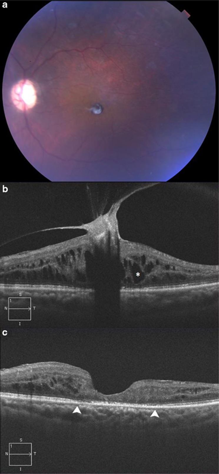
Figure 1.
(a) Color photograph shows a small, darkly pigmented, well-circumscribed lesion involving the central macula with an overlying membrane. (b) OCT shows a hyperreflective preretinal lesion in the fovea with associated VMT distorting the macular architecture. The retinal features underlying the lesion cannot be ascertained owing to severe shadowing artifact. Cystoid macular edema (*) and retinal thickening are also evident. The increased reflectivity of the lesion's anterior surface and deep underlying shadowing are characteristic of congenital simple hamartoma of the retinal pigment epithelium. (c) Optical coherence tomography shows restored foveal anatomy and resolved VMT following pars plana vitrectomy and tumor excision. There is focal disruption of the photoreceptors and retinal pigment epithelium (between arrowheads).

