
Figure 1.
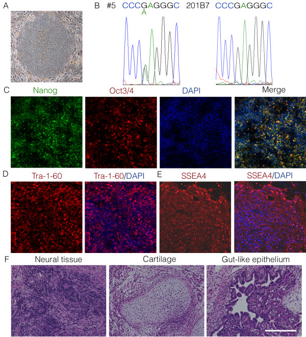
RP patient’s iPSCs. (A) A colony of RP#5 iPSCs derived from an RP patient’s skin cells. (B) DNA analysis of the rhodopsin gene in the RP#5 iPSCs and control 201B7 iPSCs. (C-E) Immunostaining for pluripotent markers; the nuclei were counterstained with DAPI (blue). (C) Nanog and Oct3/4. (D) Tra-1-60. (E) SSEA4. (F) Teratoma formation assay showing that the RP#5 iPSCs gave rise to all three germ layers, confirming their pluripotency. Scale bar, 1 mm (A), 250 μm (C-F).