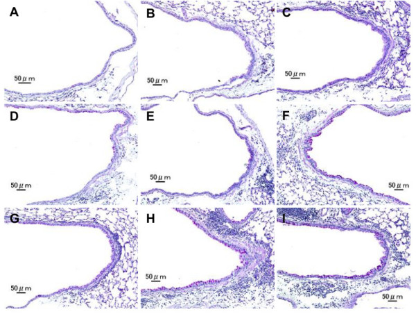
Figure 4.

Effects of test samples on pathological changes in the lungs. No pathologic alterations were seen in the lungs of the control (A). Slight bronchitis with slight proliferation of goblet cells that have mucus stained pink with PAS was seen in airway epithelium and slight infiltration of inflammatory cells in the submucosa of airways exposed to H-ASD + LPS 1 (B). Bronchitis with slight proliferation of goblet cells and moderate infiltration of inflammatory cells such as neutrophils and lymphocytes were seen in the submucosa of airways exposed to H-ASD + LPS 10 (C). Very slight proliferation of goblet cells in the airway epithelium and very slight infiltration of inflammatory cells such as lymphocytes were seen in the submucosa of airways exposed to OVA alone (D). The pathological alteration in the airway epithelium and the submucosa of airways exposed to OVA + LPS 1 (E) were almost the same. The pathological changes exposed to OVA + LPS 10 (F) were somewhat stronger than those of OVA + LPS 1. Slight goblet cell proliferation and mild infiltration of inflammatory cells were found in the submucosa of airways exposed to H-ASD + OVA (G). Moderate goblet cell proliferation, accumulation of numerous inflammatory cells in the submucosa of airway, and fibrous thickening of the subepithelial layer were seen in the airways exposed to H-ASD + OVA + LPS 1 (H) and moderate goblet cell proliferation, severe infiltration of inflammatory cells in the submucosa of airways exposed to H-ASD + OVA + LPS 10 (I). (A–I) PAS stain; bar = 50 μm.
