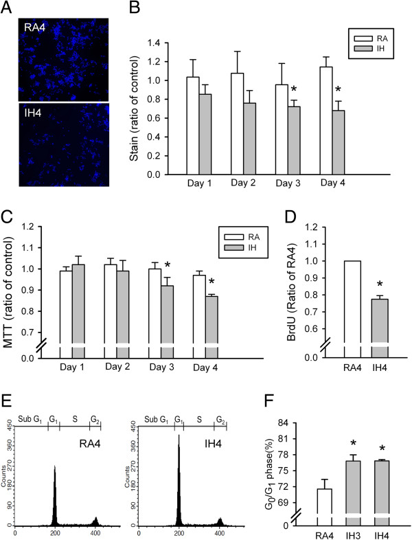
Figure 3.

Intermittent hypoxia (IH) effects on PC12 cell numbers, cell viability, cell proliferation and cell cycle progression. (A) Numbers of PC12 cells as evaluated using Hoechst staining (blue) and confocal microscopy. (B) Quantitative PC12 cell numbers after exposure to normoxia (RA) and IH for 1–4 days (n = 5 for per group). (C) PC12 cell numbers as determined by MTT assay after exposure to RA and IH for 1–4 days (n = 5 for per group). (D) PC12 cell proliferation determined by BrdU cell proliferation ELISA assay kit after exposure to 4 days of RA (RA4, n = 8) and IH (IH4, n = 8). (E) PC12 cell cycle progression after exposure to RA4 (n = 8), IH3 (n = 7) and IH4 (n = 7) as evaluated by propidium iodide staining and flow cytometry. Percentages of cells in G0/G1 phase arrest (E and F). *p < 0.05 compared with RA in (B) and (C) or RA4 in (D) and (F). Values are means ± SEMs.
