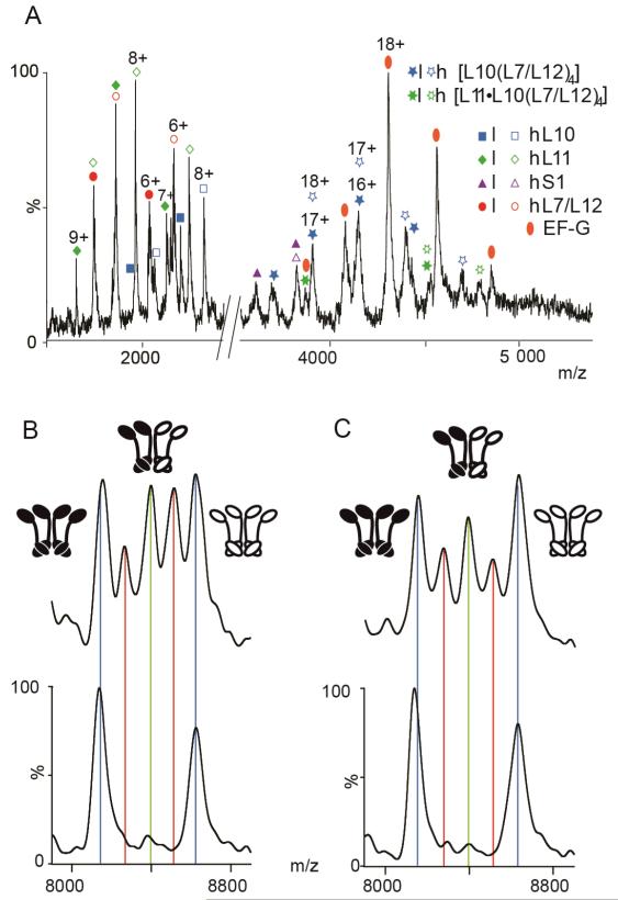
Figure 3.
L7/L12 exchange from ribosomes trapped in the posttranslocational state. A) Low m/z region of a mass spectrum recorded for an equimolar solution of l- and h-ribosome-EF–G complex trapped in the posttranslocational state by fusidic acid, prior to subunit exchange. B–C) The 6+ charge state of (L7/L12)4 stripped complex at time t=0 (lower spectra) and after 3h incubation at 37°C (upper spectra) for ribosomes in: B) the absence of EF–G and C) for the EF–G complex. Peaks corresponding to tetramers with different ratios of (l:h) L7/L12 are highlighted in blue (4:0/0:4), red (3:1/1:3) and green (2:2).

