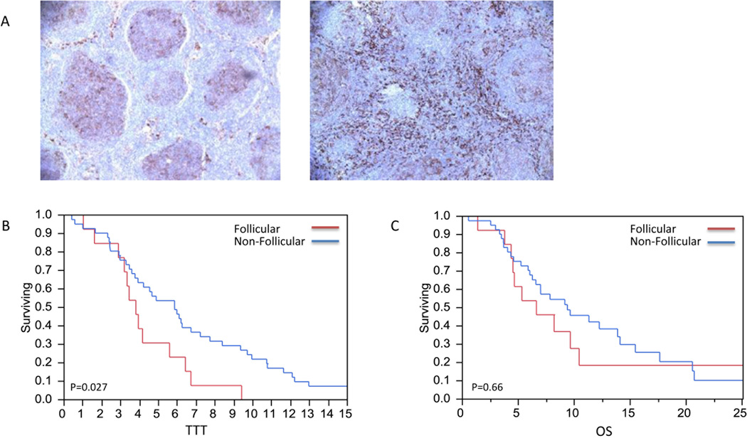
Figure 1. CD14+ cells pattern of expression and TTT.
A) Follicular and non-follicular pattern of CD14+ staining. B) CD14+ cells localized to the follicle had a shorter time to transformation compared to those that were not localized to the follicle (2.8 vs 5.9 years, p=0.27). C) Follicular CD14+ cells were not associated with improvement in OS (p=0.66).

