Figure 1.
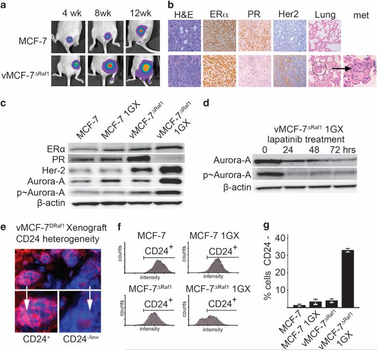
Establishment of MCF-7 and vMCF-7ΔRaf-1 breast cancer xenografts. (a) Tumor xenografts imaging in live animals of MCF-7 (upper row) and vMCF-7ΔRaf-1 (lower row) expressing the firefly luciferase reporter lentivector at 4, 8 and 12 weeks after mammary fat pad injection. (b) Paraffin sections of xenograft tumors (12 weeks) showing: hematoxylin and eosin (H&E) staining of low-grade tubular tumors for MCF-7 (upper row) and high-grade vMCF-7ΔRaf-1 tumors (lower row); expression of ER in both xenografts; loss of progesterone receptor (PR) and HER-2/Neu expression in vMCF-7ΔRaf-1 xenografts; and H&E staining of lungs showing development of metastases in vMCF-7Raf-1 xenografts. (c) Immunoblot analysis of parental and cancer cells re-cultured from tumor xenografts (1GX) showing that vMCF-7ΔRaf-1 1GX cells retain the expression of ER, lack expression of progesterone receptor and overexpress HER-2/Neu and Aurora-A. (d) Immunoblot analysis of vMCF-7ΔRaf-1 1GX cells treated with 1 μM lapatinib showing reduced expression of total and p-Aurora-A. (e) Immunofluorescence analysis showing tumor cell heterogeneity for the luminal marker CD24 in vMCF-7ΔRaf-1 xenografts. CD24 receptor was labeled in red and DNA was labeled in blue with Hoechst dye. (f) FACS analysis showing that only vMCF-7ΔRaf-1 1GX cells developed a subpopulation of CD24–/low cells (~30%), while MCF-7, MCF-7 1GX and vMCF-7ΔRaf-1 displayed a CD24+ phenotype. (g) Graph showing the percentage of MCF-7 and variant cells displaying a CD24–/low phenotype from three independent experiments (±s.d.).

