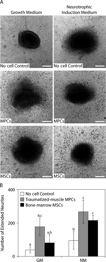
Figure 6.
Neurotrophic activity of MPCs and MSCs in DRG co-culture assays. (A) The DRGs were imaged using ×4 phase-contrast microscopy after co-culture with MPCs or MSCs for 3 days in growth medium or neurotrophic induction medium; scale bar = 250 µm. (B) The number of neurites that extended beyond the average neurite length in DRG medium (1.75 mm) as a result of co-culture with MPCs or MSCs in growth medium or neurotrophic induction media; a,b,cp < 0.05, one-way ANOVA with SNK post hoc comparisons and n = 4

