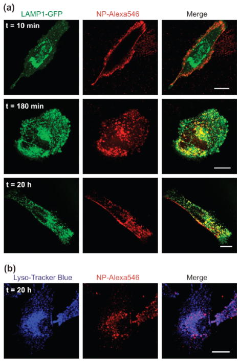
FIGURE 3.
Characterization of the vesicles containing NPs after endocytosis. (a) Confocal micrographs of LN-229 cells transiently transfected with the lysosomal marker, LAMP1-GFP, following incubation with PEI–PEG functionalized NPs. Images show representative cells 10 min (top), 3 h (middle), and 20 h (bottom) after incubation at 37 °C. (b) Confocal micrographs of LN-229 cells after overnight endocytosis of streptavidin functionalized NPs via surface biotinylation. The endolysosomal compartment was stained with LysoTracker Blue. All scale bars are 20 μm.

