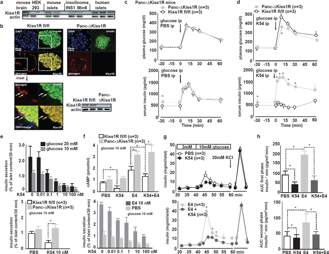
Figure 4. Kisspeptin1 at nanomolar concentrations inhibits GSIS in a Kiss1R-dependent manner. Pancreas Kiss1R is located in β-cells.
A Representative IB for Kiss1R mouse brain, HEK 293T cells, mouse islets, INS1 rat insulinoma cells, Min6 mouse insulinoma cells and human islets. Mouse brain, mouse islets, insulinoma cells and human islets express Kiss1R. HEK 293T cells do not expess Kiss1R.
B (left) Immunohistochemistry for insulin, glucagon and Kiss1R in pancreas from Kiss1Rfl/fl mice. Kiss1R immunoreactivity colocalizes with insulin-positive β-cells but not with glucagon-positive α-cells. 20× magnification. Pseudocoloring: red: glucagon, green: insulin, yellow: Kiss1R, blue: nucleus counter-stain with DAPI (left bottom) inset of previous image at 40× magnification.
(right top) Immunohistochemistry for insulin, glucagon and Kiss1R in pancreas from Panc-ΔKiss1R mice. Kiss1R immunoreactivity is lacking in Panc-ΔKiss1R islet.
(right bottom) Representative islet IB from Kiss1Rfl/fl and Panc-ΔKiss1R mice. Kiss1R is absent in Panc-ΔKiss1R islets.
C ipGTT in Kiss1Rfl/fl and Panc-ΔKiss1R mice during ip co-injection of PBS and glucose. (top) GT is similar in Kiss1Rfl/fl and Panc-ΔKiss1R mice. (bottom) baseline fasting glucose is slightly elevated in Panc-ΔKiss1R mice as compared to Kiss1Rfl/fl littermates. In vivo GSIS is similar in Kiss1Rfl/fl and Panc-ΔKiss1R mice (mean±SEM, *P<0.05).
D ipGTT in Kiss1Rfl/fl and Panc-ΔKiss1R mice during ip co-injection of 10 nM K54 and glucose. K54 impairs GT (top) and GSIS (bottom) in Kiss1Rfl/fl but not in Panc-ΔKiss1R mice (mean±SEM, *P<0.05).
E (top) Dose response curve of K54 and K10 inhibition of GSIS from WT mouse islets during static at 10 or 20 mM glucose. Both K54 and K10 inhibit GSIS in a dose-dependent manner from 0 to 100 nM at both 10 and 20 mM glucose; (bottom) GSIS from Kiss1R fl/fl and Panc-ΔKiss1R islets treated with PBS K10 or K54. K10 or K54 (both 10 nM) inhibit GSIS from Kiss1R fl/fl but not from Panc-ΔKiss1R islets.
F cAMP synthesis and GSIS in response to K54 and to incretin analogue exendin-4 (E4) in Kiss1R fl/fl and Panc-ΔKiss1R islets. (top) K54 impairs cAMP synthesis in Kiss1Rfl/fl but not in Panc-ΔKiss1R islets. E4 stimulates cAMP synthesis similarly in both Kiss1Rfl/fl and in Panc-ΔKiss1R islets. K10 reduces E4-stimulated cAMP levels in Kiss1Rfl/fl but not in Panc-ΔKiss1R islets. (bottom) During static incubation of mouse islets, K54 impairs GSIS and also E4 potentiation of GSIS from islets in a dose dependent manner (mean±SEM, *P<0.05).
G Islet perifusion assay in WT islets in response to K54 and to E4. (top) K54 impairs both first and second phasea of GSIS and (bottom) E4-potentiated first and second phase GSIS; End of perifusion shows similar insulin release upon KCL induced depolarization (mean±SEM, *P<0.05).
H Area under the curve (AUC) of (top) first and (bottom) second phase GSIS from WT mouse islets treated with PBS, K54, E4 or K54+E4. K54 inhibits both first and second phases of GSIS and E4 potentiated GSIS (mean±SEM, *P<0.05).

