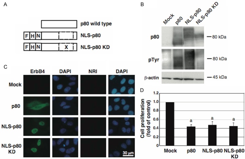
Figure 2.

Effects of forced expression of the p80 wild type and mutants on their autophosphorylation and subcellular localization and on cell proliferation. A: The schematic structures of p80, NLS-p80, and NLS-p80 KD. F, a Flag tag; H, a HA tag; N, a NLS; X, a point mutation substituting Lys of residue 751 to Arg; a dashed box, the tyrosine kinase region. B-D: Cells were transfected with each of the expression plasmids for p80, NLS-p80, and NLS-p80 KD, or the empty vector (Mock), and analyzed by Western blotting for p80, phosphorylated tyrosine residues (pTyr), and β-actin (B), by immunofluorescence staining for p80 (green) (C), and by cell proliferation assay (D). C: Cells were incubated with an antibody against ErbB4 or normal rabbit immunoglobulin (NRI), and counterstained with DAPI (blue). D: Data are presented as mean ± S.E. (fold of control) of relative values normalized to the mean value of the control cells, obtained from three independent experiments. a, p < 0.01.
