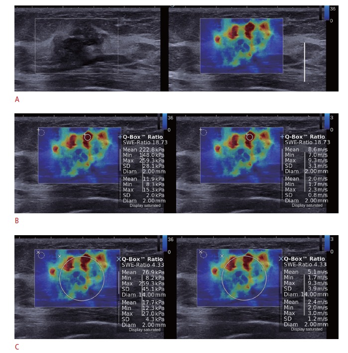
Fig. 1. B-mode and shear-wave elastography (SWE) images in a 46-year-old woman with a pathologically proven invasive ductal carcinoma.

A. SWE (right) and B-mode images (left) on split-screen mode show a 20-mm, irregular, spiculated mass with red, heterogeneous elasticity. B. The mean, maximum, and standard deviation of elasticity values were measured in kPa (left) and in m/sec (right) by placing the region-ofinterest (ROI) over the stiffest part of the lesion. C. The ROI for measuring the elasticity value (wSD) was placed to include the whole breast lesion and the stiffest part of the lesion.
