
Fig. 1. Ultrasonography (US) images in a 40-year-old woman who underwent a routine check-up.
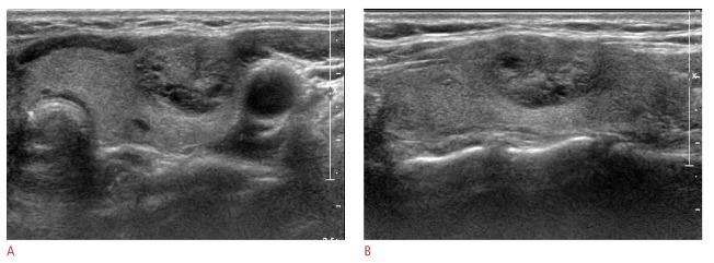
A 18-mm thyroid mass in the left thyroid gland shows mixed isoechogenicity, a well-defined margin, and parallel orientation without suspicious features on US (A, transverse image; B, longitudinal image). Thyroid Imaging Reporting and Data System category 3 and a total risk score of 0 were assigned. This thyroid nodule was diagnosed as negative for malignancy on cytologic examination and showed no change at follow-up US.