
Fig. 2. Ultrasonography (US) images in a 58-year-old woman undergoing a routine examination.
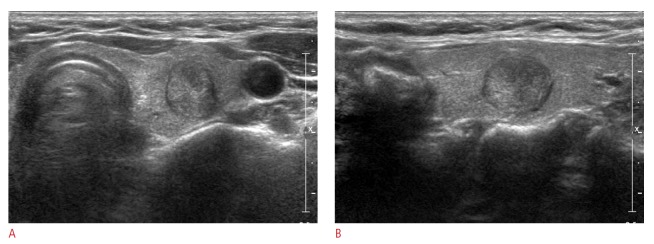
A 14-mm thyroid mass in the right thyroid gland shows two suspicious US features: complete solid and taller-than-wide shape on US (A, transverse image; B, longitudinal image). Thyroid Imaging Reporting and Data System category 4b and a total risk score of 1 were assigned. This thyroid nodule was diagnosed as malignant on cytological examination and proved to be papillary thyroid cancer on surgery.