
Fig. 3. Ultrasonography (US) images in a 25-year-old woman who underwent a routine check-up.
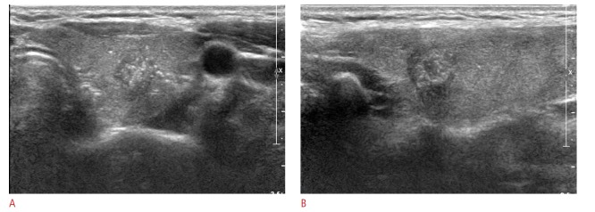
A 10-mm thyroid mass in the right thyroid gland is seen to be a complete solid with hypoechogenicity, an irregular margin, and a taller-thanwide shape on US (A. transverse image; B. longitudinal image). Thyroid Imaging Reporting and Data System category 5 and a total risk score of 10 were assigned. This thyroid nodule was diagnosed as malignant on cytological examination and proved to be papillary thyroid cancer on surgery.