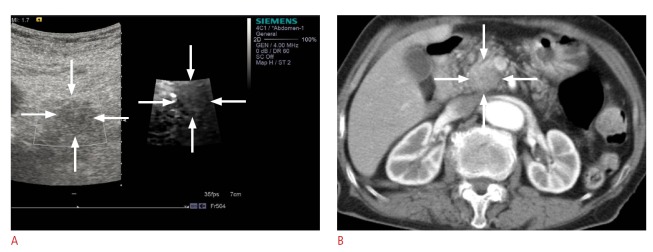
Fig. 3. A 62-year-old woman with focal pancreatitis.

A. On a conventional B-mode image, the lesion appears as a hypoechoic mass (left image). On a virtual touch tissue imaging elastographic image, the lesion shows a stiffness similar to the surrounding pancreatic parenchyma (echogenicity score, 3) (right image). The shear wave velocity of the lesion and surrounding pancreatic parenchyma was 2.08 m/sec and 2.02 m/sec, respectively. B. Axial computed tomography scan shows an ill-defined hypodense mass in the pancreatic head region.
