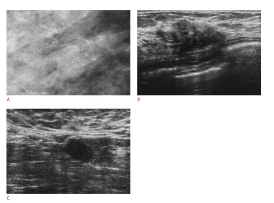
Figure 1. A 42-year-old woman with a palpable mass in her right breast which proved to be an invasive ductal carcinoma.

A. Mammogram demonstrates suspicious microcalcifications in the right breast. B. Breast ultrasonography reveals a 3 cm, irregularly-shaped and microlobulated margined hypoechoic mass with echogenic foci within the right upper outer quadrant. C. Abnormal lymph nodes without fatty hilum in the right axilla can be seen. The patient underwent an ultrasound-guided 14-gauge core needle biopsy with a ductal carcinoma in situ identified based on the subsequent histology. Invasive carcinoma was found following surgical excision with the presence of metastatic lymph nodes detected after axillary lymph node dissection.
