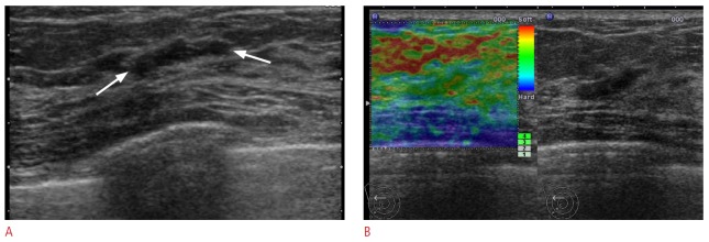
Figure 1. A 50-year-old woman with a Breast Imaging Reporting and Data System (BI-RADS) category 3 lesion on supplemental screening ultrasonography.

A. Transverse B-mode ultrasonogram shows an 11-mm, oval, circumscribed mass (arrows). B. Ultrasound (US) elastographic image shows the entire mass as green, indicating an elasticity score of 1. US-guided core biopsy revealed a fibrocystic change. Thirty-six month follow-up ultrasonography showed the lesion was not changed (not shown).
