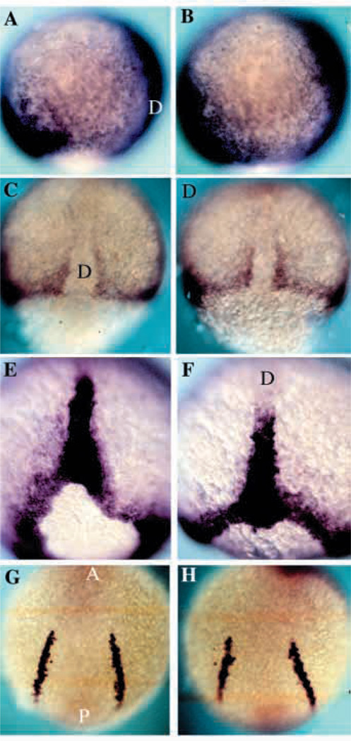
Fig. 7.
DV patterning is normal in nrd. Expression analysis of the mesodermal genes BMP-4, Snail1, Brachyury and SCL in wild-type (A,C,E,G) and nrd (B,D,F,H) embryos at 80–90% epiboly to 5 somite stage. (A,B) BMP-4 is expressed on the ventral side of the gastrula and the leading edge of the invagingating tissue on the dorsal side in both wild-type and nrd embryos. Anterior top and dorsal to the right. (C,D) Dorsal view of the expression of snail1 as seen by in situ hybridization shows the normal paraxial mesoderm in nrd (anterior to the top). (E,F) Zebrafish Brachyury is expressed in a ring around the blastopore and in the forming notochord at the end of gastrulation. nrd embryos show a normal pattern of expression of Brachyury (F) as compared to wild type (E). Dorsal view, anterior to the top in both E and F. (G,H) Expression of SCl in the blood precursors at the 5 somite stage shows normal development of a mesoderm derivative, blood, at a slightly later stage in an nrd (H) embryo compared to a wild type (G). This region is just ventral to the neural crest forming region. (Note: nrd embryos presented in this figure were from nrd mutant clutches in which homozygous were not distinguishable from heterozygous siblings) D, dorsal; A, anterior; P, posterior.

