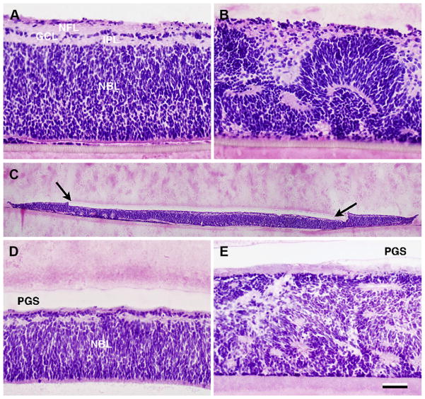
Fig. 4.
Donor tissue after 7–9 days in vitro. Hematoxylin and eosin staining. A–B: E40 neuroretinal explants without addition of PGS membrane. In A, a well laminated explant is seen with a neuroblastic cell layer (NBL) consisting of multiple rows of undifferentiated cells, a thin inner plexiform layer (IPL), a ganglion cell layer (GCL) with 1 row of cells and a nerve fiber layer NFL in the innermost part. In B severe folding of the retinal architecture is seen in another explant. C–E: PGS-Retina explants: In C, the full extent of the composite explant with the Laminin-PCL-PGS membrane (between arrows) attached to the inner retina can be seen. D: Detail. The explanted retina displays an NBL and rudimentary inner layers. E: Rosetted explant without any apparent lamination. Scale bar = 50 μm (A–B and D–E) and 500 μm (C).

