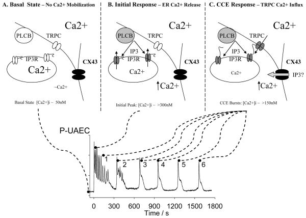
Figure 2. Proposed mechanisms underlying the [Ca2+]i response to ATP over 30 minutes in P-UAEC.
Upper Panels A–C: Note in all upper panels the darker the shading the more active/open the protein. Lower Graph: This represents a typical [Ca2+]i response to 100uM ATP in a single P-UAEC cell over 30 minutes. A) In the basal state the intracellular Ca2+ pool in the Endoplasmic Reticulum (ER) is full and the IP3Rs are at rest. PLC-beta is inactive due to the lack of agonist stimulation. The TRPC3 channels are closed for lack of stimulation. Nonetheless CX43 channels are maintained in a permanently open state. B) On initial agonist stimulation the initial upslope of the first increase in [Ca2+]i is solely due to release of intracellular ER pool of Ca2+. This is in turn due to PLC-beta activation and the resulting rise in Ins(1,4,5,)P3 which then activates IP3R. C) Once the ER is empty of Ca2+ (at the peak) the IP3R detects this change and in turn undergoes a physical change in shape, so allowing physical communication with overlying TRPC channels in the plasma membrane and they are triggered to open. This in turn allows in Ca2+ from the extracellular space. Initially this serves to extend the first major peak. We suspect the rapid superimposed oscillations are the result of rapid [Ca2+]i sensitive changes in the activity of Ins(1,4,5)P3-3 kinase. As in most cells the system operating to produce the first major peak of [Ca2+]i then shuts itself off through as yet unidentified mechanisms. Thereafter, over the remaining 25 minutes, the TRPC activation system again activates periodically, suggesting IP3 production is maintained and the ER remains partially or nearly empty, or other regulatory mechanisms now apply to TRPC activation. While the source of Ca2+ for burst 2 onwards is essentially extracellular through TRPC channels, the process remains entirely dependent on CX43 remaining open to communicate between the cells. In this figure we have shown Ins(1,4,5)P3 moving between the cells (IP3) which is certainly possible but equally CX43 may communicated by passing Ca2+ or providing electrical coupling. Further studies are needed on this specific point.

