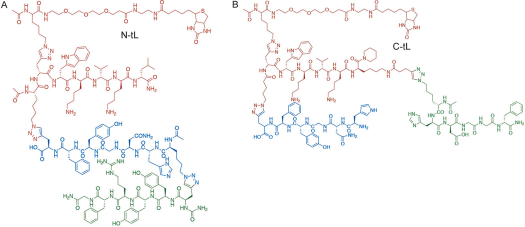
Figure 1.
Molecular structures of the N-tL (A) and C-tL (B) developed against the C-terminal epitope of Akt2 near the pS474 residue. The 1° and 2° ligand branches are common to both PCC agents, and are drawn in red and blue, respectively. The poly (ethylene glycol)-linked biotin groups were included in the development process and thus do not represent interfering perturbations.

