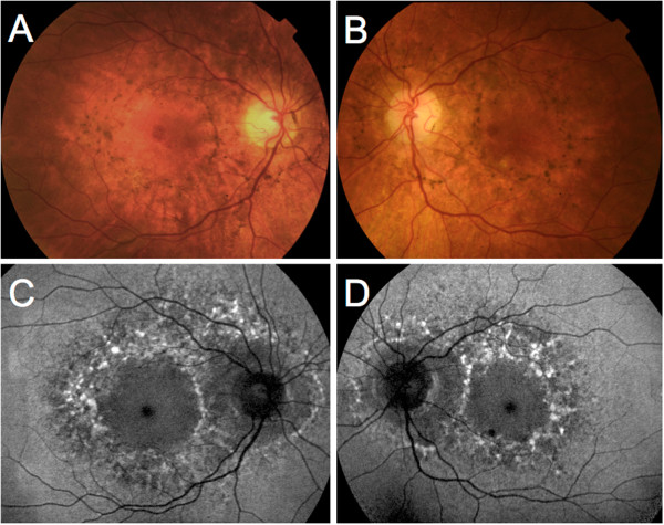
Figure 1.

Color fundus photograph and fundus autofluorescence of Patient 1. A, B, Color fundus photograph of patient 1. Note the hyperpigmented lesions surrounding the macula and the optic disc, associated with depigmentation areas of the retinal pigment epithelium. C, D, Fundus autofluorescence of the same patient. The hyperpigmented spots correspond to mainly increased autofluorescence, whereas the hypopigmented spots correspond a decreased granular autofluorescence signal.
