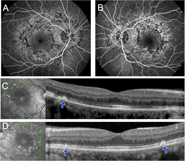
Figure 2.

Fluorescein angiography and SD-OCT of Patient 1. A, B, Fluorescein angiography showed blockage of background fluorescence in the areas of hyperpigmentation and hyperfluorescence due to retinal pigment epithelium window defects in the depigmented areas. C, D, On SD-OCT scans taken through the planes indicated by the green arrows, hyperpigmented areas corresponded to a hyperreflective dome-shaped lesion that seemed to originate from the retinal pigment epithelium (blue arrows).
