
FIGURE 6.
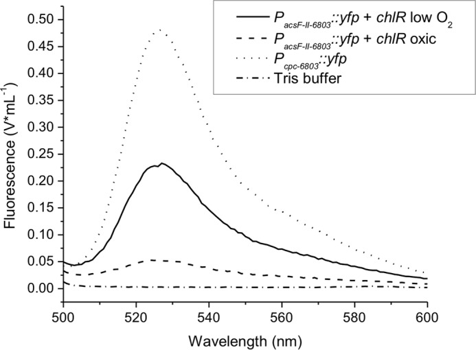
YFP expression in E. coli under control of the acsFII promoter from Synechocystis 6803 and NStrepChlR from Synechococcus 7002. YFP expression was detected and quantified as fluorescence for E. coli BL21(DE3) cells that carried both plasmids pLM7 and pLM5 and that were grown under microoxic conditions or oxic conditions. E. coli cells (strain TOP10F′) with plasmid pAQ1Ex::PcpcBA::yfp grown under oxic conditions served as the positive control. Cells were harvested by centrifugation, washed in 50 mm Tris-HCl buffer at pH 8.0, resuspended in the same buffer, and adjusted to an OD600 nm of 0.5 for the measurements. Cell suspensions were incubated at room temperature and exposed to air; measurements were taken at several times until the YFP fluorescence signal developed fully (data not shown). The fluorescence spectra showing the maximum signal for all condition are as follows: PcpcBA-6803::yfp (pAQ1Ex::PcpcBA::yfp) (positive control; dotted line), PacsF-II-6803::yfp (pLM7) and NStrepChlR (pLM5) microoxic sample (solid line), PacsF-II-6803::yfp and NStrepChlR oxic conditions (dashed line), and Tris-HCl buffer (negative control; dash/dotted line).