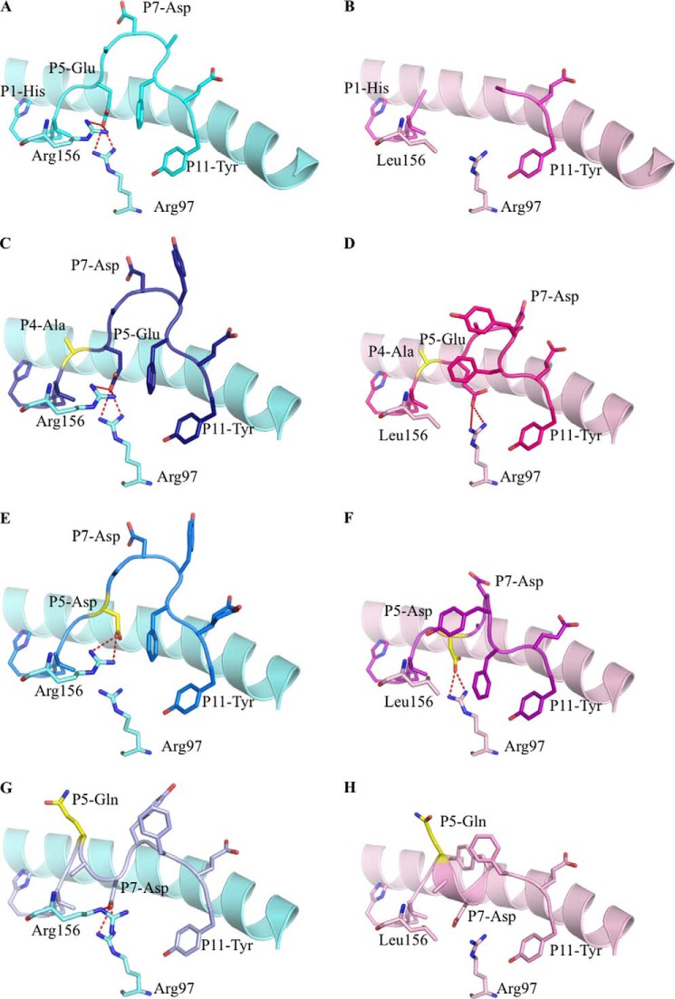
FIGURE 4.
Binary structures of pHLA complexes. Structures of four HPVG epitopes (stick format) bound to HLA-B*35:08 (pale blue) or HLA-B*35:01 (pale pink) (25). The HPVG peptide is in cyan and pink in panels A and B, HPVG-A4 is in dark blue and magenta in panels C and D, the HPVG-D5 is in blue and purple in panels E and F, and the HPVG-Q5 is in pale blue and pale pink in panels G and H bound to HLA-B*35:08 and HLA-B*35:01, respectively. Arg-97 and the polymorphic residue 156 (Arg in HLA-B*35:08 and Leu in HLA-B*35:01) of the antigen binding cleft are represented in stick and colored accordingly to the HLA. The variations in the HPVG peptide are highlighted in yellow at positions 4 and 5. The red dashed lines represent the salt bridge between the P5 or P7 of the peptide with the Arg-97 and Arg-156.

