FIGURE 3.
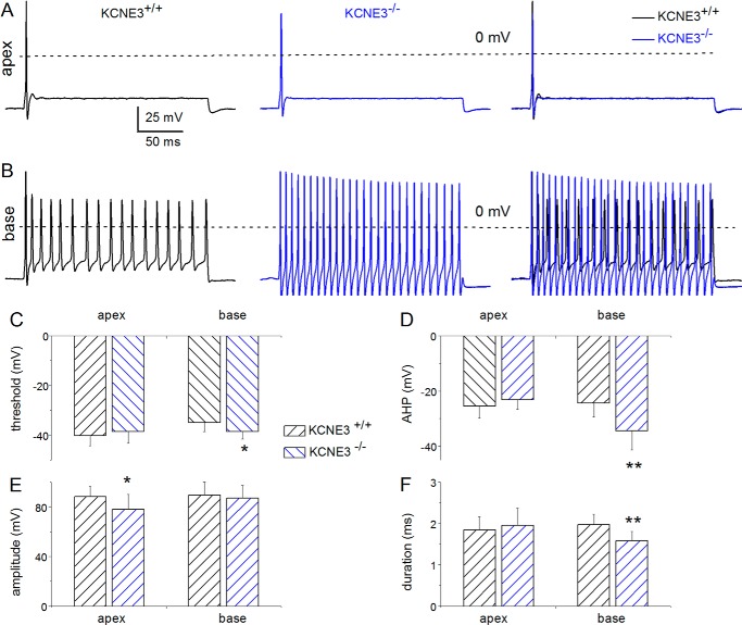
Membrane properties of Kcne3+/+versus Kcne3−/− SGNs at P12, the genesis of hearing. A, assessment of the evoked P12 SGN APs from rapidly adapting neurons isolated from the apical aspects of the cochlea from the Kcne3+/+ (left panel) and Kcne3−/− mice (middle panel) and superimposed traces (right panel). The recording conditions were identical in the wild-type and null mutant neurons. APs were evoked by injecting 0.1 nA current for ∼250 ms. B, representative recordings from slowly adapting SGNs from the base of the cochlea showing APs from Kcne3+/+(left panel) and Kcne3−/− (middle panel) neurons. We superimposed traces (right panel) for comparison. C–F, summary data from analyses of AP properties. C, threshold voltage; D, extent of AHP; E, AP amplitude, and F, AP duration. Significant differences are noted, where *, p < 0.05; **, p < 0.01. Also see Table 1 where the number of samples is listed.

