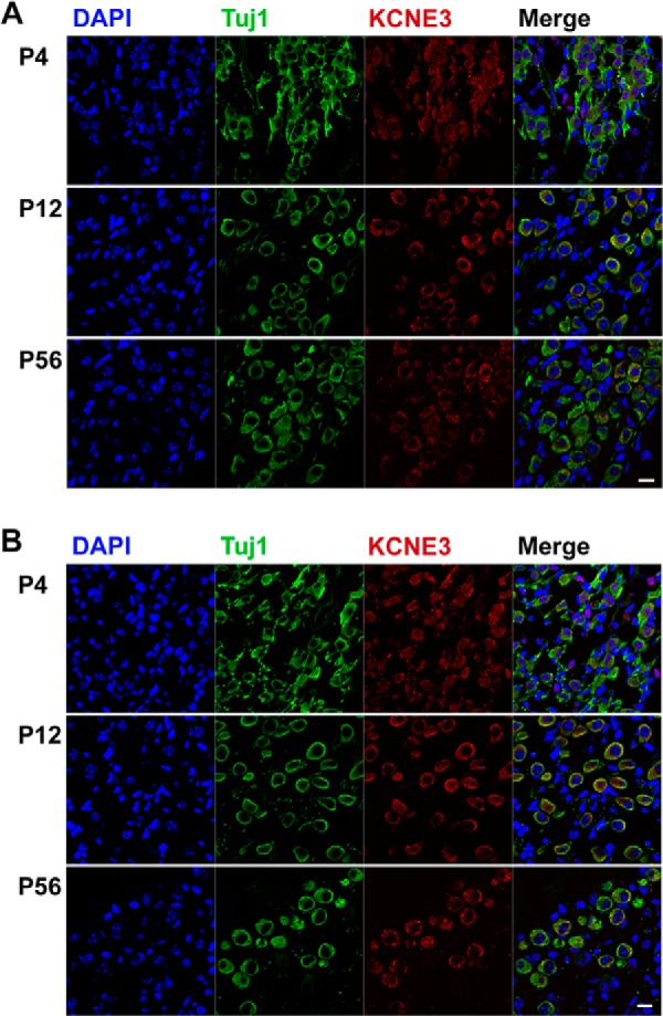
FIGURE 7.

Identification of chronological expression of KCNE3 in SGNs. A, cryosections of P4 and P12 and 2 m (8 weeks (wk) or P56) apical SGNs were stained with an antibody directed against KCNE3 (in red), and neurons were identified with positive reactivity toward Tuj1, the neuronal marker. The nuclei were stained with DAPI. The expression of KCNE3 was seen from P4 to P56. B, similar observation was made for basal SGNs. These findings were observed from more than 200 sections from three preparations of each age. Scale bar, 20 μm.
