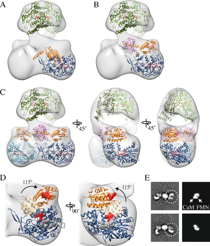
FIGURE 7.
Molecular model of the nNOS-CaM holoenzyme identifying a rotation of the FMN domain to a deshielded state. A, fit of the reductase domain (PDB ID: 1TLL) showing the FMN domain (orange) in the shielded arrangement with the aligned CaM binding helix (PDB ID: 3HR4) and NADPH-FAD domains (blue). B, fit of the reductase domain with CaM (pink) and showing the rotation of the FMN domain around the flexible hinge (*) to a proposed FMN-released, deshielded state. Cofactors are shown in red. C, complete molecular model the nNOS-CaM complex in the deshielded state. D, close-up view of the reductase domain with the FMN in the shielded (light orange) and deshielded (dark orange) states indicating a 115° rotation. E, comparison of two-dimensional averages of uncross-linked nNOS-CaM (left panels) and two-dimensional projections of the proposed reductase-CaM deshielded arrangement (right panels).

