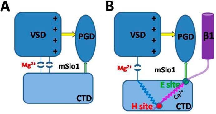
FIGURE 7.

A hypothetical scheme for mechanism of the calcium- and magnesium- and voltage-dependent activation in BK(β1) channels. The rounded rectangles denote the VSD (dark blue) and CTD (light blue) of mSlo1 (A) and mSlo1-β1 (B), respectively. The blue oval denotes the PGD. The purple cylinder denotes the transmembrane domain of β1, and the light purple curve denotes the N terminus of β1. Yellow and green arrows and pink and blue springs denote the possible interaction pathways. The H site serves as a fixed pulley (red circle) transferring the force coming from CTD to enlarge the distance between VSD and CTD to reduce the Mg2+ sensitivity, and the E site (green circle) then optimizes the direction of the gating force coming from the Ca2+ binding sites.
