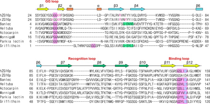
FIGURE 2.
Sequence alignment of human ZG16p, rat ZG16p, and Jacalin-related mannose binding-type lectins. Pink boxes indicate residues involved in mannose binding at the first sugar-binding site. Proposed heparin-binding residues (Lys33, Lys36, Arg37, Arg55, and Arg58 in rat ZG16p) and conserved residues in human ZG16p (Lys36, Arg37, and Arg55) are shown in purple. Second binding site in Banlec and Griffithsin is shown in light green. Asp151 in ZG16p and corresponding aspartic acid residues in other lectins are colored in cyan. Secondary structure of human ZG16p is shown above the amino acid sequence. Sequences are aligned with MATRAS (52) and CLUSTAL W (53).

